

On this page
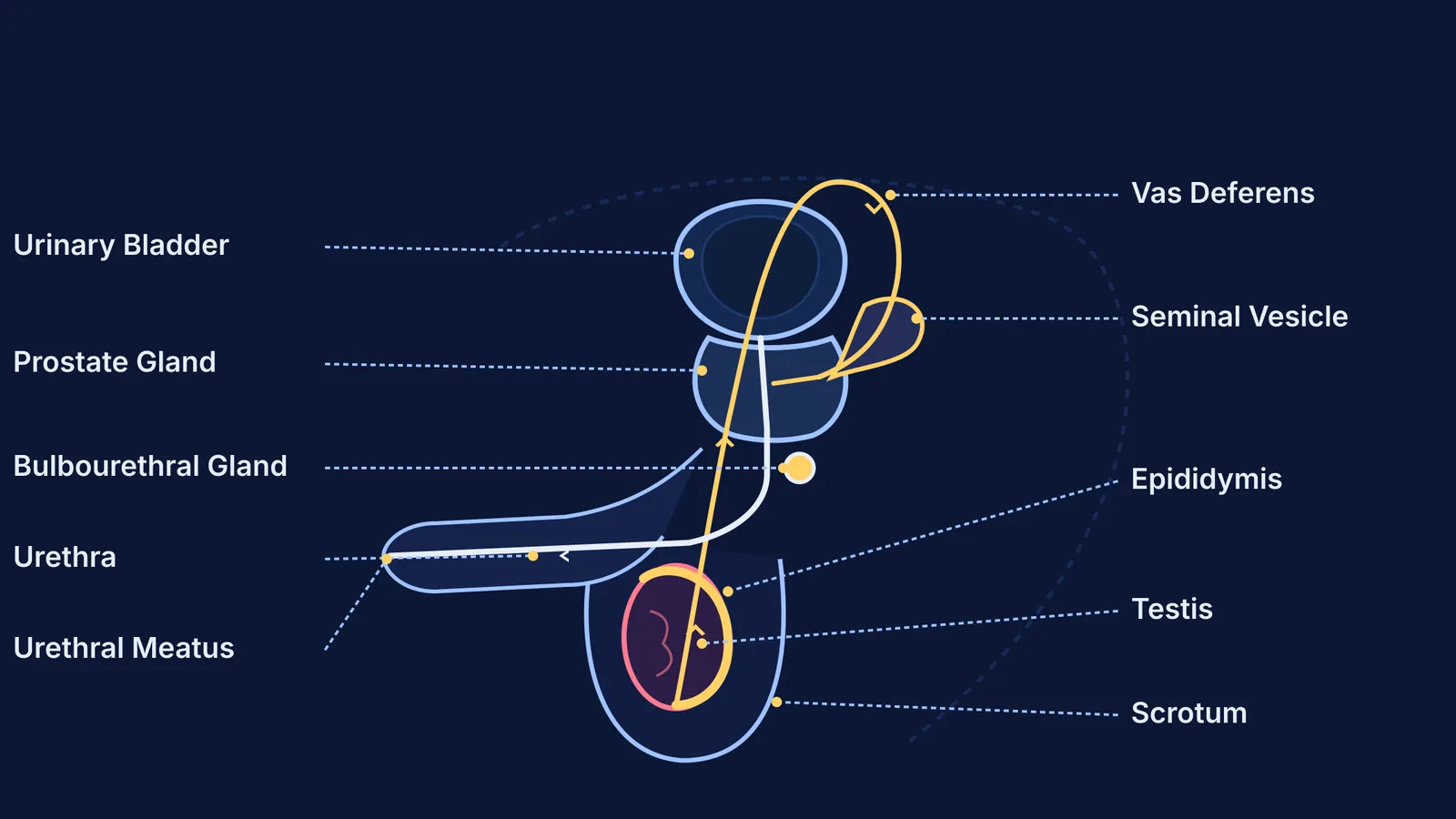
In board examinations, biology diagrams test functional anatomy rather than artistic drawing. An accurate sketch of the male reproductive system demonstrates the route sperm follow from production to ejaculation. Examiners award marks for an unbroken pathway: primary sex organs in the scrotum, accessory storage ducts looping into the pelvis, tributary glands adding nutritive secretions, and a shared urogenital exit.
What is the correct anatomical order to label the male reproductive system?
Examiners trace diagrams following the sequence of male sex accessory ducts set out in the NCERT textbook. Spermatogenesis begins within the seminiferous tubules of the testis, passing into the rete testis and through delicate vasa efferentia into the epididymis. The epididymis caps the posterior border of each testis, serving as the site for sperm maturation and temporary storage. According to clinical duct transit research by Mavis J. Rowley and colleagues in Fertility and Sterility37502-1), spermatozoa spend 1 to 21 days (averaging approximately 12 days) traversing human male ducts.
From the tail of the epididymis, sperm enter the vas deferens, a muscular duct measuring approximately 30 cm long as noted in anatomical records on the male reproductive system. The vas deferens ascends through the scrotal neck, traverses the inguinal canal, and loops over the urinary bladder. Behind the bladder, its terminal ampulla joins the seminal vesicle duct to form the ejaculatory duct, which enters the prostate gland and joins the urethra. Our Class 10 revision guide on how organisms reproduce covers how these early concepts build toward senior exams. When memorising how separate organs connect feels abstract, the Fermi Concept Library covers every concept in biology, each with its own lesson and its place in the map.
Why are the testes located outside the abdominal cavity in the scrotum?
A classic question in CBSE and NEET asks why the human testes hang externally rather than remaining inside the abdomen. The scrotum acts as a thermoregulatory pouch maintaining testicular temperature 2 to 2.5 °C below core body temperature, an absolute physiological prerequisite for viable spermatogenesis confirmed in the NCERT textbook. Each testis contains approximately 250 testicular lobules, each housing 1 to 3 coiled seminiferous tubules.
External positioning protects temperature-sensitive germ cells from abdominal heat. The scrotal wall adjusts its surface area through involuntary contraction and relaxation of the dartos and cremaster muscles, as documented in anatomical studies of the male reproductive system. The surrounding pampiniform venous plexus provides counter-current cooling, lowering arterial blood temperature before it reaches developing sperm.
How do accessory glands contribute to semen along the sperm pathway?
Sperm cells constitute only 2% to 5% of total ejaculate volume; the remainder is seminal plasma produced by three accessory glands. Quantitative analysis by Derek H. Owen and David F. Katz in the Journal of Andrology demonstrates that paired seminal vesicles contribute 65% to 75% of semen volume. This alkaline fluid contains fructose to energise sperm motility, prostaglandins to stimulate reproductive tract contractions, and clotting proteins that temporarily coagulate semen.
Beneath the bladder, the chestnut-shaped prostate gland surrounds the first segment of the urethra. As established in the Journal of Andrology, prostatic fluid accounts for 20% to 30% of semen volume, supplying citric acid, calcium, and proteolytic enzymes that liquefy the coagulum. Below the prostate, paired bulbourethral glands contribute under 5% of volume, releasing alkaline mucus that lubricates the urethra and neutralises residual acidic urine prior to ejaculation, as catalogued in the WHO Laboratory Manual for the Examination and Processing of Human Semen. This preparation equips sperm for the journey described in our guide on the pathway from fertilisation to implantation.
What common diagram mistakes cause students to lose marks in board exams?
Examiners routinely penalise five avoidable errors: confusing ureters with the vas deferens, placing the prostate above the bladder, omitting the epididymis, drawing the vas deferens cutting straight through the bladder cavity, and omitting the external urethral meatus.
To secure full marks under timed conditions, follow this five-step sequence:
Sketch the oval urinary bladder and pelvic outline lightly to set proportions.
Place the scrotal sac and oval testis inferiorly, adding the crescent epididymis along the posterior border.
Draw the vas deferens looping over the bladder dome before descending into an expanded ampulla.
Position the seminal vesicle behind the bladder base, add the prostate around the bladder neck, and place the bulbourethral gland below.
Extend the urethra through the penile shaft to the urethral meatus, aligning straight label lines horizontally with a ruler.
For more step-by-step sketching guides across physics, chemistry, and biology, review our collection of essential CBSE Class 10 science diagrams. When solving multi-step questions under exam conditions, the Fermi Problem Library provides worked problems with a step-by-step solver that explains the reasoning rather than printing an answer.


