

On this page
In short
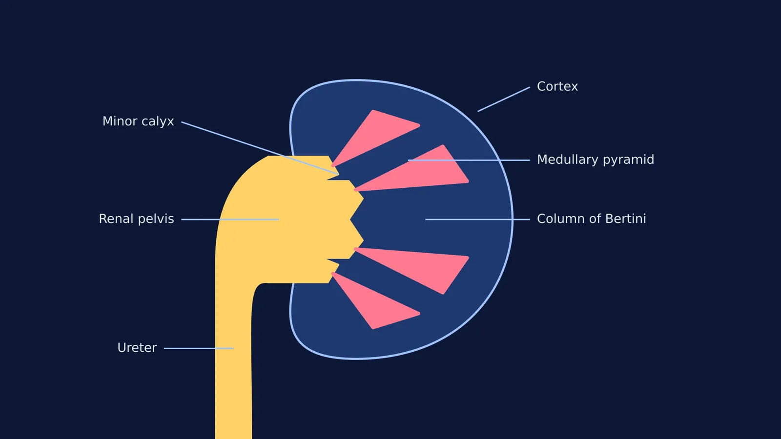
The longitudinal section of an adult human kidney reveals an outer cortex and an inner medulla divided into 8 to 18 medullary pyramids. These pyramids drain into calyces, which merge into the funnel-like renal pelvis leading to the ureter.
When you study the human excretory system, understanding the longitudinal section of the kidney is essential. This view reveals the internal machinery that filters blood and concentrates urine. In adult humans, each kidney is roughly 12 cm long and weighs up to 190 g (Wikipedia). Slicing it lengthwise exposes two main zones: an outer cortex and an inner medulla, which feed into a central drainage system.
What Are the Renal Cortex and the Columns of Bertini?
The renal cortex is the outer layer of the kidney's tissue (Wikipedia). It has a granular appearance because it contains the renal corpuscles—the microscopic structures where blood filtration begins. Each corpuscle consists of a glomerulus and Bowman's capsule, which work together to filter out wastes, extra salts, and water from the bloodstream.
The cortex is not just a uniform outer rim. Projections of cortical tissue extend inward, separating the structures of the medulla. These extensions are called the columns of Bertini, or renal columns (Wikipedia). They help anchor the cortex and provide a pathway for blood vessels to reach the outer layers of the organ. These columns also provide structural support, keeping the kidney's internal architecture stable.
How Many Pyramids Are in the Renal Medulla?
Beneath the cortex lies the renal medulla, the inner part of the kidney. The medulla is organized into distinct, cone-shaped masses known as renal pyramids. A typical human kidney contains between 8 and 18 of these pyramids (Wikipedia).
The pyramids appear striped because they contain the straight segments of renal tubules and collecting ducts, which are responsible for concentrating urine. Their broad bases face the cortex, while their pointed tips, called renal papillae, project inward toward the center of the kidney. Each human kidney contains approximately one million nephrons (Wikipedia), and while their filtering heads sit in the cortex, their loops of Henle dip deep into these medullary pyramids to process the filtrate.
If you are trying to picture how these structures fit together in three dimensions, the Fermi Concept Library can help. It covers every concept in biology, providing each with its own lesson and showing its place in the map of human anatomy.
What Is the Function of the Renal Pelvis?
Once urine is concentrated in the medullary pyramids, it must be collected and transported out of the body.
Minor Calyces: The tip of each pyramid rests inside a small, cup-like structure called a minor calyx.
Major Calyces: Several minor calyces merge to form larger spaces known as major calyces.
Renal Pelvis: The renal pelvis is a funnel-like dilated part of the ureter inside the kidney (Wikipedia). It acts as a central collection point where the major calyces empty urine before it leaves the organ.
Ureter: The renal pelvis tapers as it exits the kidney through a central notch called the hilum, continuing downwards as the ureter. The hilum is the anatomical gateway for the kidney; alongside the ureter, the renal artery and renal vein also pass through this opening.
How Do You Draw the Longitudinal Section for Exams?
Anatomy exams frequently ask students to draw and label the longitudinal section of a kidney. Memorising a visual sequence makes this much easier. Practicing this diagram is one of the highest-yield activities for biology students because the structure perfectly dictates its function.
Draw the outline: Start with a large bean shape, making sure to include an inward notch (the hilum) on the concave side.
Sketch the pelvis: Draw a funnel shape emerging from the hilum, branching into two or three major calyces, which then split into smaller minor calyces.
Add the pyramids: Draw 6 to 8 triangular shapes (representing the 8 to 18 actual pyramids, Wikipedia). Rest the tip of each triangle in a minor calyx.
Label the cortex and columns: The space between the outside edge and the pyramids is the cortex. Label the tissue dipping between the triangles as the columns of Bertini.
When revising these diagrams, Fermi's AI tutor can assist. Instead of just printing an answer, it holds a line of reasoning across a session. It can work out what you already know and build a plan for the session, drawing the figure and checking your understanding as it goes.
For more on the excretory system, you can explore Ammonotelic, ureotelic and uricotelic animals: why waste differs and Regulating kidney function: ADH, the RAAS and ANF explained. To read about the scientists behind these discoveries, see William Bowman and the capsule that filters blood in the kidney.


