

On this page
Polysaccharides are macromolecular chains of monosaccharide units joined by glycosidic bonds. In secondary-school curricula and competitive exams, students frequently compare carbohydrates by their structure and biological role. While starch and cellulose are familiar examples of glucose polymers, inulin and chitin illustrate how specialised monomer chemistries create distinct storage and structural functions.
What is inulin and how is it structured?
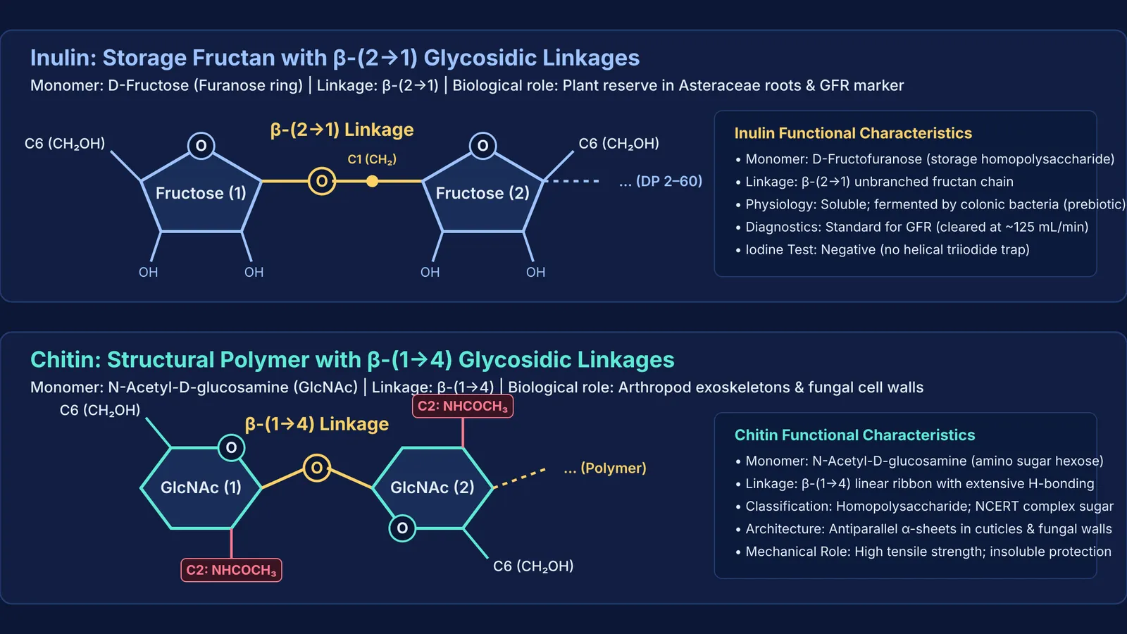
Inulin is a storage fructan composed of repeating D-fructofuranose units connected by β-(2→1) glycosidic linkages, typically ending with a single α-D-glucopyranosyl residue. In a review in The Journal of Nutrition, Kathy Niness showed that native chicory inulin has a degree of polymerisation spanning 2 to 60 units, with an average chain length between 10 and 20 fructose residues.
Because human digestive enzymes cleave only α-glycosidic bonds, inulin passes unhydrolysed through the stomach and small intestine into the colon. As noted by Marcel Roberfroid in Critical Reviews in Food Science and Nutrition, colonic bacteria ferment inulin into short-chain fatty acids, making it a functional dietary prebiotic. In plants, members of the Asteraceae family—such as dahlia (*Dahlia variabilis*) and chicory—store inulin in roots and tubers instead of starch.
Why is inulin used to measure kidney filtration?
In human physiology, inulin is the clinical reference standard for measuring glomerular filtration rate (GFR). In their 1935 study in the Journal of Clinical Investigation, James Shannon and Homer Smith demonstrated that inulin satisfies every requirement of an ideal filtration marker.
When injected intravenously, inulin filters freely across glomerular capillaries due to its modest size and neutral charge. Renal tubule cells neither reabsorb nor secrete it. Consequently, the volume of plasma cleared of inulin per minute equals the true filtration rate, averaging 125 millilitres per minute in healthy adults.
If you are connecting physiological filtration to carbohydrate structure, the Fermi Concept Library covers every concept in physics, chemistry, biology, mathematics and computing, each with its own lesson and its place in the map.
What is chitin and where is it found in nature?
Chitin is the second most abundant natural biopolymer after cellulose. It is an unbranched homopolysaccharide of N-acetyl-D-glucosamine (GlcNAc) units linked by β-(1→4) glycosidic bonds. Each GlcNAc unit is an amino sugar where the carbon-2 hydroxyl group is replaced by an acetamido group (-NH-CO-CH3).
Chitin provides structural protection across two major groups:
Arthropod exoskeletons: As reviewed by Hans Merzendorfer and Lars Zimoch in the Journal of Experimental Biology, chitin microfibrils assemble into antiparallel sheets in insect and crustacean cuticles, embedded within sclerotin and hardened in crabs by calcium carbonate.
Fungal cell walls: As shown by Neil Gow, Jean-Paul Latgé, and Carol Munro in Microbiology Spectrum, chitin forms an inner scaffold cross-linked to β-(1,3)-glucans that withstands osmotic turgor pressure.
Why is chitin called a complex polysaccharide if it is a homopolymer?
Students studying NCERT Class 11 Biology often notice that the textbook groups chitin under complex polysaccharides while biochemical sources call it a homopolymer. Both descriptions are correct:
It is a homopolymer because it contains only one repeating monosaccharide species: N-acetyl-D-glucosamine. Unlike heteropolysaccharides such as hyaluronic acid, it has no alternating sugar partners.
It is considered complex because its building block is a chemically modified amino sugar rather than a simple unmodified hexose.
Exam questions regularly test this distinction. To explore how structural and storage products fit into plant and animal metabolism, see our guide on primary and secondary metabolites in NCERT and our overview of biomolecules in chemistry.
How do you distinguish between inulin, chitin, and related polysaccharides?
Use this three-step diagnostic checklist to classify polysaccharides quickly:
Monomer type: Fructose indicates inulin; α-D-glucose indicates starch or glycogen; β-D-glucose indicates cellulose; N-acetyl-D-glucosamine indicates chitin.
Glycosidic linkage: β-(2→1) bonds define inulin; β-(1→4) bonds build the flat, linear ribbons of cellulose and chitin; α-(1→4) and α-(1→6) bonds form the coiled helices of starch and glycogen.
Iodine test: Starch forms a helical inclusion complex with triiodide ions, turning intense blue. Inulin, cellulose, and chitin lack this helical geometry and give a negative test. To understand mutarotation and reducing properties, consult our reducing and non-reducing sugars guide.
When solving carbohydrate classification numericals or GFR clearance problems, the Fermi Problem Library provides worked problems with a step-by-step solver that explains the reasoning rather than printing an answer.


