

On this page
How do pressure gradients drive breathing?
The process of breathing, or pulmonary ventilation, depends on a simple rule of physics: Boyle's Law. As the volume of a closed space increases, the pressure inside it drops. The human chest cavity operates exactly on this principle 1.
When the muscles of the chest wall and the floor of the thorax change the volume of the chest cavity, they create pressure gradients between the air inside the lungs (intra-alveolar pressure) and the air outside the body (atmospheric pressure). Because air always flows down its pressure gradient, it will naturally move from an area of higher pressure to an area of lower pressure until the two pressures are equal.
The lungs themselves are completely passive during this process. They do not have skeletal muscle tissue of their own to force expansion or contraction. Instead, they rely on the adhesive nature of the pleural fluid, which effectively glues the outside of the lungs to the inside of the thoracic wall 1. When the chest wall moves, the lungs are pulled along with it.
What happens during inspiration?
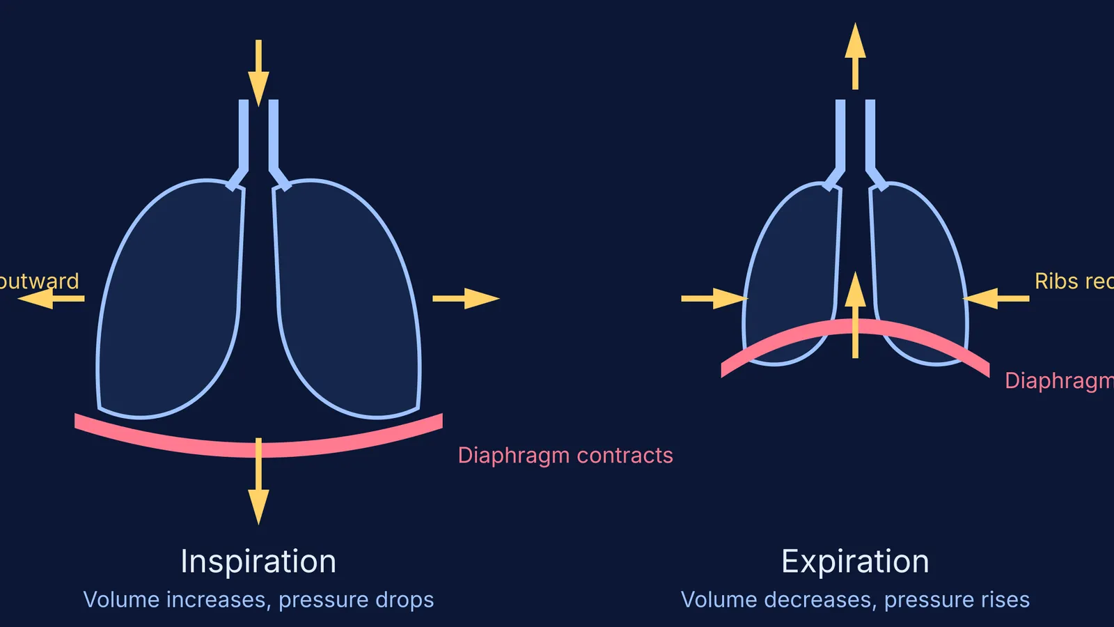
Inspiration, or inhalation, is an active process that requires energy to contract muscle fibres. It begins with the contraction of the primary muscle of inspiration, the diaphragm. When the diaphragm contracts, it moves inferiorly—flattening downwards toward the abdominal cavity. This movement significantly increases the vertical length of the thoracic cavity 1.
At the same time, the external intercostal muscles, which are located between the ribs, contract. This action lifts the ribs upward and outward, expanding the rib cage's diameter. The combined effect of the diaphragm dropping and the ribs lifting is a large increase in thoracic volume.
Because the pleural fluid keeps the lungs attached to the chest wall, the internal volume of the lungs increases as well. Following Boyle's Law, this expansion causes the intra-alveolar pressure to drop below atmospheric pressure. The resulting pressure gradient creates a vacuum effect that draws air into the lungs from the outside environment. During a normal, quiet breath, these pressure gradients move about 500 millilitres of air (known as the tidal volume) into the lungs 1.
Depending on the body's needs, inspiration can be driven more by one muscle group than the other. A deep breath, known as diaphragmatic breathing, relies heavily on the contraction of the diaphragm. A shallow breath, known as costal breathing, relies more on the contraction of the external intercostal muscles 1.
Why is quiet expiration a passive process?
Quiet breathing, also known as eupnea, is the normal breathing you do while resting. During eupnea, expiration is a completely passive process that does not require any active muscle contraction or cognitive thought 1.
To breathe out, the muscles that caused inspiration simply relax. The diaphragm returns to its original dome-like shape, moving upward into the chest cavity. The external intercostal muscles relax, allowing the rib cage to drop back down and inward due to the natural pull of gravity. Furthermore, the lung tissue and the thoracic wall are highly elastic, meaning they naturally recoil to their smaller resting size once the stretching force is removed.
As the chest and lungs shrink, their internal volume decreases. This compresses the air inside, causing the intra-alveolar pressure to rise slightly above atmospheric pressure. Consequently, air is pushed out of the lungs into the atmosphere.
If you are trying to understand the sequence of events from muscle contraction to volume and pressure changes, the Fermi Concept Library has every concept in physics, chemistry, biology, mathematics and computing, each with its own lesson and its place in the map. Tracing the cause-and-effect chain is a reliable way to avoid confusing the roles of different muscles during exams.
Which muscles control forced breathing?
While quiet expiration is passive, forced breathing—known as hyperpnea—is a highly active process. When you exercise heavily, sing, or blow out candles, your body needs to move larger volumes of air more rapidly. This requires active muscle manipulation for both inspiration and expiration 1.
During forced inspiration, the body must expand the chest cavity even further than usual. To achieve this, accessory muscles of the neck, including the scalenes, contract alongside the diaphragm and external intercostals. These neck muscles actively lift the thoracic wall, creating a greater increase in lung volume and a deeper pressure gradient to draw in more air.
Forced expiration is similarly active. To expel air quickly, the body recruits accessory muscles to forcefully compress the chest cavity. The abdominal muscles, such as the obliques, contract to push the abdominal organs upward against the diaphragm, driving it further into the thorax 1. Simultaneously, the internal intercostal muscles contract, actively pulling the ribs downward and inward to rapidly squeeze the rib cage.
This rapid, forceful reduction in thoracic volume creates a much steeper pressure gradient, driving air out of the lungs quickly and completely.
Understanding these mechanics is a foundation for other physiological processes; for instance, the oxygen you breathe in is eventually used at a cellular level, as seen when exploring respiration as an amphibolic pathway. Similarly, the way oxygen is processed in different organisms can be seen when looking at respiration in plants.


