

On this page
After a ligation, some plasmids carry your gene, many have closed up empty, and most bacteria usually take up nothing at all. Cloning works only if you can tell these three kinds of cell apart on a plate. Two textbook tricks do it, both resting on one idea.
For the full chapter, see our guide to restriction enzymes, PCR and cloning vectors for NEET and the methods for getting DNA into host cells.
What is insertional inactivation?
Insertional inactivation is the inactivation of a gene by inserting a fragment of DNA into the middle of its coding sequence, so the cell loses the trait the gene gave it (Wikipedia, insertional inactivation). Cloning vectors exploit this on purpose: in pBR322, the restriction sites inside the antibiotic-resistance genes allow for insertional inactivation (Wikipedia, pBR322). The vector therefore carries two kinds of marker. A selectable marker lets only transformed cells grow. A second marker, with the cloning site inside it, reports whether the insert went in.
How does pBR322 show which colonies are recombinant?
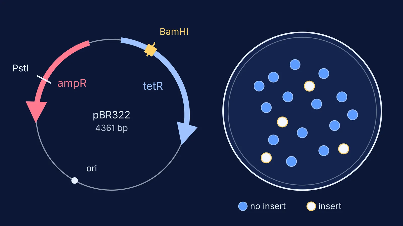
pBR322 is a 4,361 base-pair E. coli plasmid built in 1977 in Herbert Boyer's lab and named after Francisco Bolívar and Raymond Rodriguez (Wikipedia, pBR322; original paper: Bolivar et al., Gene, 197790000-2)). It carries resistance genes for ampicillin and tetracycline (Addgene vector database).
In the GenBank sequence of pBR322, the tetracycline-resistance gene runs from base 86 to 1276, the ampicillin-resistance gene (bla) from 3293 to 4153, and the only BamHI site sits at base 375, inside the tet gene (NCBI GenBank J01749). The single PstI site lies inside the ampicillin gene (Addgene vector database).
So if you cut with BamHI and ligate in your gene:
Untransformed cells: die on ampicillin and on tetracycline.
Cells with an empty, re-closed pBR322: grow on both.
Recombinant cells: grow on ampicillin, die on tetracycline.
Because the BamHI site sits inside the tet gene, an insert there gives an ampicillin-resistant, tetracycline-sensitive cell, and you find it by replica plating from an ampicillin plate onto a tetracycline plate. Our post on Esther Lederberg and replica plating explains that copying step. Recombinants are found by what they fail to do.
How does blue-white screening identify recombinant colonies?
The pUC plasmids of Jeffrey Vieira and Joachim Messing were the first plasmids built to use this colour screen (Wikipedia, blue-white screen; paper: Vieira and Messing, Gene, 198290015-4)). The vector carries lacZα, a short piece of the β-galactosidase gene, with the multiple cloning site inside it.
The host strain carries lacZΔM15, a version of the gene missing amino acids 11 to 41 (Wikipedia, blue-white screen). Neither fragment works alone, but together they form a working enzyme, which is called α-complementation (GoldBio).
The plate contains X-gal, a colourless lactose look-alike. Working β-galactosidase splits it, and the product dimerises into an insoluble blue pigment, 5,5'-dibromo-4,4'-dichloro-indigo (Wikipedia, blue-white screen). IPTG, an inducer of the lac operon, is often added to the plate too: lacZα is under lac control, so IPTG switches its expression on (GoldBio).
An insert in the cloning site breaks lacZα, so no enzyme forms, X-gal stays colourless and the colony is white; an empty vector gives a blue colony (Addgene blog). Unlike pBR322, you read the answer on the first plate.
Why do you still need an antibiotic on the plate?
X-gal alone cannot tell a white recombinant from a white cell with no plasmid, because a colony with no vector at all is also white (Wikipedia, blue-white screen). The antibiotic in the plate does that job; Addgene's guide, for example, plates transformations on ampicillin plates containing X-gal (Addgene blog). Three things can still mislead you:
Satellite colonies: cells with no vector can grow once the antibiotic around a resistant colony has been used up (Wikipedia, blue-white screen).
Blue recombinants: an insert that is in frame with lacZα and has no stop codon can leave the colony blue (Wikipedia, blue-white screen).
White does not prove the right insert, so labs confirm picks by colony PCR, restriction digest or sequencing (Addgene blog).
Worked example: reading a plate in four steps
Say a pUC-type vector cut in its cloning site, mixed with your gene and ligase, then transformed and spread on ampicillin + X-gal + IPTG. Next morning you see 40 blue and 12 white colonies.
Ask what the antibiotic removed: apart from satellites, every colony here has a plasmid, because cells without one could not grow on ampicillin.
Read the colour: the 40 blue colonies have an intact lacZα, so they are almost certainly empty vector.
Pick white: the 12 white colonies are likely recombinants; pick big, well-separated ones, not tiny satellites.
Confirm: check a few picks by colony PCR or a digest before you trust them.
If the difference between selection and screening still feels slippery, the Fermi Concept Library gives each concept its own lesson and shows where it sits in the map. For practice, the Fermi Problem Library has worked problems with a step-by-step solver that explains the reasoning rather than printing an answer.


