

On this page
If you are studying plant anatomy, you will quickly encounter the three main tissue systems: dermal, vascular, and ground. While the dermal system forms the outer skin and the vascular system acts as the plumbing, everything else falls into the ground tissue category.
What is the ground tissue system in plants?
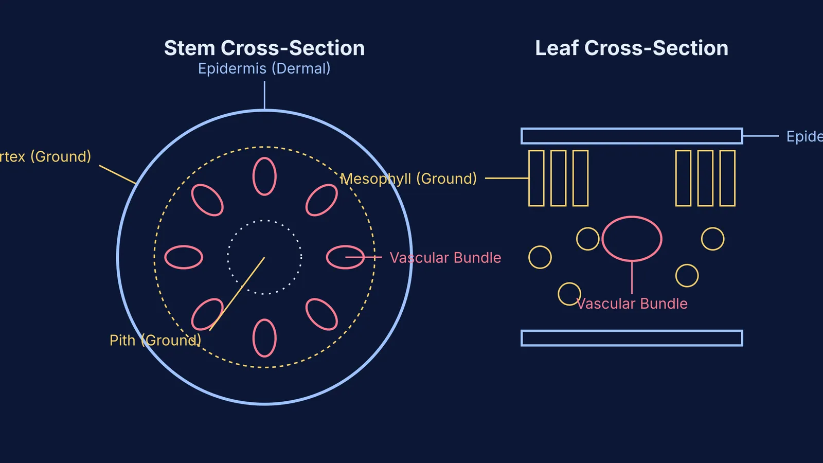
By definition, the ground tissue system of plants includes all tissues that are neither dermal nor vascular (Wikipedia). It makes up the bulk of the plant's body and is responsible for a variety of essential functions, including structural support, metabolic activity, and the storage of food and water.
You can think of it as the packing material that fills the spaces between the outer skin (epidermis) and the internal transport tubes (xylem and phloem). But it is far from inactive. In leaves, for example, the ground tissue is the primary site of photosynthesis.
What tissues are included in the ground tissue system?
When you look at a cross-section of a stem or root under a microscope, the ground tissue system is typically divided into distinct zones. Moving from the outside in, these include:
Cortex: The outer region located just inside the epidermis.
Endodermis: Often considered the innermost layer of the cortex, it acts as a selective boundary.
Pericycle: A layer located just inside the endodermis, surrounding the vascular tissues.
Medullary Rays: Strips of tissue that extend radially between the vascular bundles, connecting the center of the stem with the outer layers.
Pith: The central core of the stem or root, primarily used for storage.
In leaves, the ground tissue is not separated into cortex and pith. Instead, it is called the mesophyll. The mesophyll contains the chloroplast-rich cells where the plant manufactures its food.
(For more on how plant tissues grow, read about meristems: apical, intercalary and lateral.)
How do ground tissue cells work together?
The regions listed above are built from three main types of simple cells, each with a specific job:
Parenchyma: These versatile cells usually remain alive at maturity and have thin primary walls. They handle photosynthesis in the mesophyll, store food and water in the cortex and pith, and help repair wounds.
Collenchyma: These cells have irregularly thickened walls and provide flexible structural support to growing plant parts. They are often found just beneath the epidermis in growing shoots and leaf stalks.
Sclerenchyma: These cells have thick, lignified secondary walls and are often dead at maturity. They provide the main stiffness and rigid structural support to the plant. According to historical uses, sclerenchyma fibers are industrially valuable for making fabrics like linen and jute (Wikipedia).
If you have gaps in your understanding of these cell types, you can explore their lessons in the Fermi Concept Library.
How to study plant cross-sections effectively?
Memorizing the layers of a plant stem or root can be confusing. Instead of just reading the list, try this active learning framework:
Draw it yourself: Start with a blank circle representing the epidermis. Work your way inward, drawing and labeling the cortex, endodermis, pericycle, vascular bundles, and pith.
Color-code the systems: Use one color for dermal tissue, a second for vascular tissue, and a third for all ground tissues. This visual separation reinforces the rule that ground tissue is simply "everything else."
Compare and contrast: Draw a dicot stem next to a dicot root. Notice how the ground tissue organization changes—for instance, how the pith might be large and distinct in one but reduced in the other.
By mapping the tissues visually, you shift from rote memorization to a structural understanding of how the plant is built. (You can also see how structure relates to function in topics like tropic movements in plants.)


