

On this page
The human excretory system removes metabolic nitrogenous wastes—chiefly urea and uric acid—from the bloodstream while regulating body water and electrolyte levels. For Class 10 students, drawing this system is one of the most reliable scoring opportunities in biology. According to curricular guidelines from CBSE Academic, board examinations frequently feature this diagram as a 3-mark or 5-mark question, testing clean line work and correct anatomical connections between the organs and systemic blood vessels.
What are the essential organs of the human excretory system?
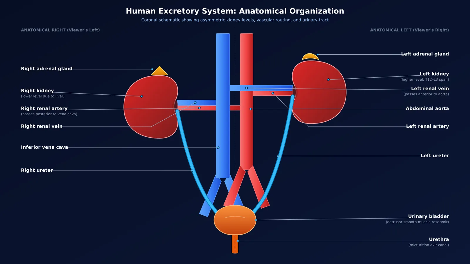
As outlined in standard medical anatomy and the NCERT Class 10 Science curriculum, the macroscopic human excretory system comprises four main organs:
A pair of kidneys: Bean-shaped organs positioned against the posterior abdominal wall on either side of the backbone. According to StatPearls anatomical guidelines, each adult kidney measures 10 to 12 cm in length, 5 to 7 cm in width, and 3 to 5 cm in thickness (often simplified to 2 to 3 cm in secondary school texts), spanning the T12 to L3 vertebrae. The medial border features a concave notch called the hilum, where vessels and the ureter connect.
A pair of ureters: Slender muscular tubes approximately 25 to 30 cm long (StatPearls). They emerge from the renal pelvis at each hilum and convey urine downward to the bladder using peristalsis.
The urinary bladder: A muscular sac situated in the pelvic cavity. Because its smooth muscle is under nervous control, it expands to store urine until pressure triggers the urge to void.
The urethra: A single canal leading from the base of the bladder to the exterior, releasing urine during micturition.
Understanding why animals excrete urea rather than ammonia clarifies why terrestrial vertebrates rely on this concentrated filtration architecture.
How should you draw the kidneys, ureters, and blood vessels step by step?
Under exam conditions, following an orderly sequence prevents messy corrections:
Draw the central blood vessels: Sketch two parallel vertical vessels down the center. The inferior vena cava sits on your left (anatomical right), and the abdominal aorta sits on your right (anatomical left).
Outline the asymmetric kidneys: Sketch two bean shapes flanking the vessels. Because the liver occupies space above the right kidney, place the right kidney roughly 1 to 2 cm lower than the left kidney (StatPearls). Ensure the concave hilum of each kidney faces inward toward the spine.
Connect the renal arteries and veins: Draw the renal arteries branching from the aorta into each hilum. Next, draw the renal veins draining from each hilum into the vena cava, showing the left renal vein crossing anterior to the aorta.
Route the ureters: Extend two smooth tubes emerging strictly from the medial hilum of each kidney, curving downward toward the pelvis.
Add the bladder and urethra: Draw the pear-shaped urinary bladder receiving the ureters at its postero-lateral base, then taper the bottom neck into the narrow urethra.
Which labelling mistakes cost marks in CBSE board exams?
Marking schemes from CBSE Academic award 1 to 2 marks for overall layout and 0.5 marks for each correct label. Evaluators regularly penalise five common mistakes:
Misplaced ureters: Drawing ureters emerging from the lower pole or outer curve of the kidney instead of the medial hilum.
Level kidneys: Drawing both kidneys at identical heights, ignoring the liver's effect on the right kidney.
Vascular mix-ups: Confusing the aorta with the vena cava, or failing to show renal vessels branching into the hilum.
Swapping bladder and urethra: Confusing the storage organ (bladder) with the exit canal (urethra).
Left-right inversion: Labelling the right side of the page as the right kidney rather than the left kidney.
Exam questions often link this macro-level diagram with the microscopic filtration described in William Bowman's discovery of the filtration capsule. To see how whole-organ anatomy connects with microscopic filtration, the Fermi Concept Library maps every concept in biology, each with its own lesson and its place in the map.
How does blood flow through the renal vessels during urine formation?
Although the kidneys account for less than 1% of body weight, they receive roughly 20% to 25% of resting cardiac output—about 1.0 to 1.2 litres of blood each minute (StatPearls).
Blood carrying urea, salts, and metabolic waste enters through the renal arteries. In the renal cortex, approximately one million nephrons filter this blood under high pressure. As detailed in the NCERT Class 10 Science textbook, the kidneys generate about 180 litres of initial filtrate daily at a glomerular filtration rate of 125 mL/min (StatPearls).
To prevent dehydration, tubular reabsorption reclaims more than 99% of this fluid along with glucose and amino acids (NCERT). The remaining 1 to 2 litres form urine, which travels through the ureters into the bladder. Purified blood exits via the renal veins into the vena cava. These balances are fine-tuned by hormonal cascades, as covered in our guide on regulating kidney function and urine concentration.
For students tackling numerical problems on filtration rates, the Fermi Problem Library provides worked problems with a step-by-step solver that explains the reasoning rather than printing an answer. For dynamic concepts that are easier to grasp in motion, the Fermi Simulation Lab offers interactive simulations for the ideas that are easier to see than to read.
What is your exam-hall diagram checklist?
Before handing in your answer sheet, verify your drawing against this quick checklist:
Right kidney placed slightly lower than the left kidney.
Both ureters and renal vessels emerge strictly from the medial hilum.
Aorta (red) and inferior vena cava (blue) positioned on the correct anatomical sides.
Urinary bladder and urethra distinctly drawn and labelled.
All label lines drawn horizontally with a ruler, never crossing one another.


