

On this page
What is the difference between euchromatin and heterochromatin?
According to the NCERT Class 12 Biology textbook, human cells package approximately 2.2 metres of DNA into an interphase nucleus typically 5 to 10 micrometres across (with typical nuclear dimensions cited at 10⁻⁶ metres). Eukaryotic cells wind the duplex around basic histone octamers to form nucleosomes—the repeating units of chromatin. Yet chromatin packaging is not uniform throughout the nucleus.
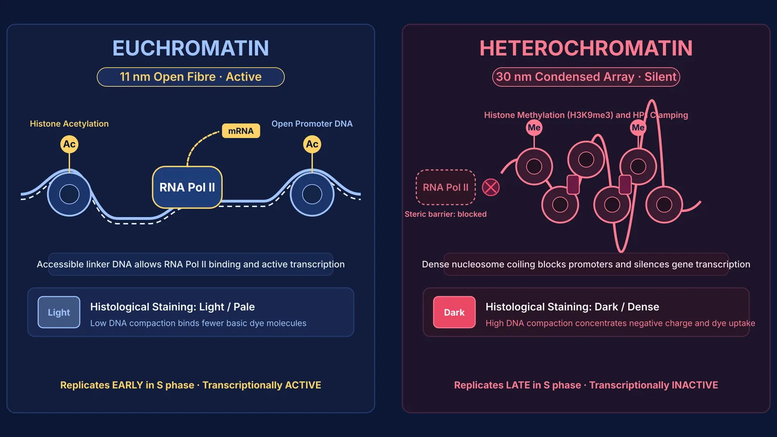
The section on Packaging of DNA Helix (Section 5.1.2 in current rationalised editions, Section 6.1.2 previously) defines the two functional states: euchromatin is loosely packed, stains lightly, and actively transcribes RNA; heterochromatin is densely packed, stains darkly, and remains transcriptionally inactive. Emil Heitz first described this distinction in 1928 after observing bryophyte (*Pellia epiphylla*) nuclei. The two states also replicate asynchronously: euchromatin replicates early in S phase, whereas heterochromatin replicates late in S phase (Lima-de-Faria, 1959). This packaging relies on the chemical properties of nucleosides and nucleotides.
Why does heterochromatin stain darkly while euchromatin stains lightly?
Under light microscopy, chromatin states are distinguished by how they take up basic histological dyes like acetocarmine, Giemsa, and basic fuchsin. This contrast reflects DNA density and chemical stoichiometry.
Because DNA's phosphate backbone carries negative charges, chromatin binds basic dye cations. In heterochromatin, tightly packed nucleosomes concentrate many base pairs into a tiny volume. This concentrated charge binds numerous dye molecules, producing high optical absorption that appears dark under magnification.
In euchromatin, the decondensed fibre spreads across a larger volume. Fewer phosphate groups occupy each unit area, binding fewer dye molecules and leaving euchromatin pale. The Feulgen reaction confirms this relationship directly (Chieco and Derenzini, 1999): acid hydrolysis unmasks deoxyribose aldehydes that react with Schiff reagent, producing deep magenta in dense heterochromatin and faint staining in euchromatin.
Why is euchromatin transcriptionally active and heterochromatin silent?
Transcriptional activity depends on steric accessibility: eukaryotic RNA polymerases and transcription factors must contact promoter DNA directly.
In euchromatin, widely spaced nucleosomes leave open linker DNA where RNA polymerase II binds promoters to initiate transcription before RNA processing and splicing. Active genes in euchromatin adopt an open conformation that is selectively digested by DNase I, whereas condensed heterochromatin resists digestion (Weintraub and Groudine, 1976).
Post-translational histone modifications control this access. Histone acetyltransferases add acetyl groups to lysine residues on histone tails, neutralizing positive charges and loosening their grip on the DNA backbone (Allfrey, Faulkner, and Mirsky, 1964). Active euchromatin exhibits high acetylation and H3K4me3. Conversely, heterochromatin is hypoacetylated and marked by H3K9me3, which recruits Heterochromatin Protein 1 to condense fibres and lock out RNA polymerase.
The Fermi Concept Library covers every concept across biology and chemistry, each with its own lesson and its place in the map, helping you connect molecular modifications to cellular traits.
What is the difference between constitutive and facultative heterochromatin?
Heterochromatin is divided into two classes based on developmental reversibility:
Constitutive heterochromatin remains permanently condensed and silent in all cells. Located at centromeres and telomeres, it consists of repetitive satellite DNA providing chromosome stability.
Facultative heterochromatin contains sequences silenced conditionally in specific lineages, retaining the ability to decondense into active euchromatin.
The classic example of facultative heterochromatin is the mammalian inactive X chromosome, described by Murray Barr and Ewart Bertram in 1949 and explained genetically by Mary Lyon in 1961 (Lyon, 1961). In female mammal cells (XX), one X chromosome condenses into a darkly staining Barr body to equalize gene dosage with males.
How to identify chromatin states in exam questions?
Exam questions mapped to the NCERT Class 12 Biology curriculum frequently test chromatin structure through comparative assertions. Use this four-step diagnostic protocol to evaluate statements:
Check physical compaction: identify whether the chromatin is an open 11-nanometre fibre (euchromatin) or a condensed 30-nanometre array (heterochromatin).
Inspect optical staining: confirm whether the region stains lightly (euchromatin) or darkly (heterochromatin) under basic histological dyes.
Verify transcriptional access: check whether the region is accessible to RNA polymerase II and sensitive to DNase I digestion (euchromatin) or sterically blocked (heterochromatin).
Distinguish reversibility: classify permanent structural silencing at centromeres as constitutive heterochromatin, and reversible developmental silencing like the Barr body as facultative heterochromatin.
Consider a problem: "A cytogeneticist observes a deeply stained peripheral body in an interphase human female nucleus that replicates late in S phase and produces no mRNA. Identify the chromatin type."
Applying the protocol, dark staining and transcriptional silence rule out euchromatin; the peripheral body in a female cell identifies the Barr body, confirming it as facultative heterochromatin.
For multi-step genetics questions, the Fermi Problem Library provides worked problems with a step-by-step solver that explains the reasoning rather than printing an answer.


