

On this page
What is the difference between endocrine and exocrine glands?
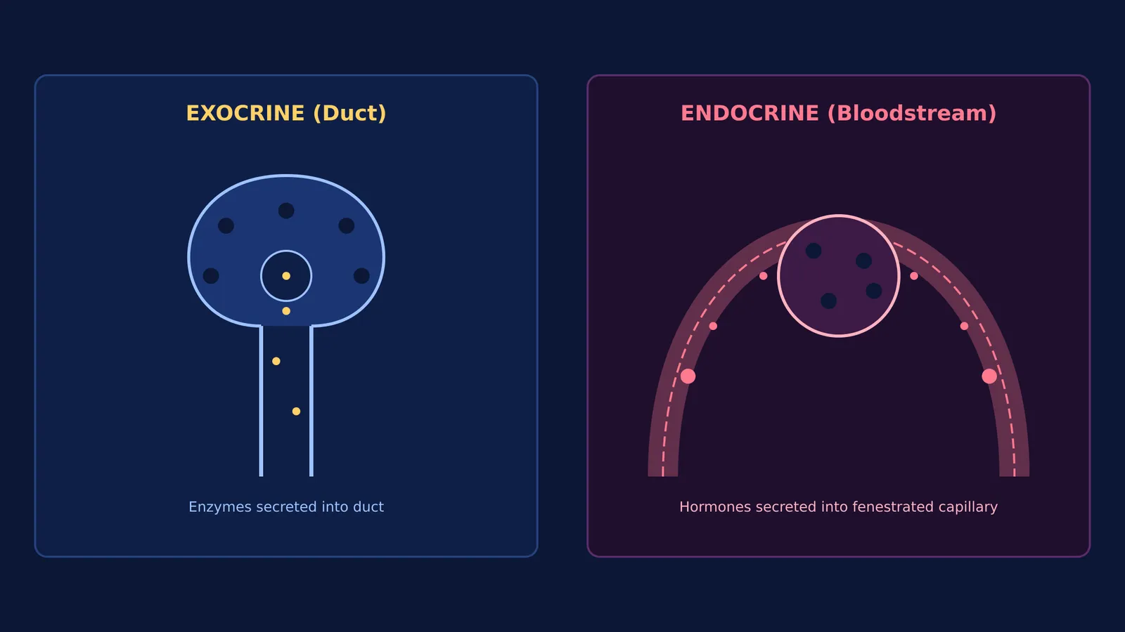
Glands are specialized epithelial structures that synthesize and secrete chemical substances. The distinction between exocrine and endocrine glands rests on their delivery architecture, as outlined in Physiology, Exocrine Gland and Physiology, Endocrine Hormones.
Exocrine glands retain ducts that route non-hormonal secretions—such as digestive enzymes, saliva, sweat, and mucus—directly onto an external or internal epithelial surface. According to Physiology, Exocrine Gland, they release substances through three cellular modes:
Merocrine: Secretion via exocytosis with no loss of cellular material, seen in salivary glands and pancreatic acini.
Apocrine: The apical portion of the cytoplasm pinches off with the secretory product, as in mammary glands.
Holocrine: The entire secretory cell ruptures and dies, as in cutaneous sebaceous glands.
Endocrine glands lose their ducts during development, making them ductless glands according to the NCERT Biology syllabus. Instead of utilizing tubes, endocrine cells secrete chemical messengers called hormones directly into interstitial fluid. These molecules diffuse into fenestrated capillaries to circulate systemically to distant target organs.
How does the pancreas perform both endocrine and exocrine functions?
Most organs belong exclusively to one category. Salivary and sweat glands are purely exocrine; the thyroid and pituitary glands are purely endocrine. The pancreas is unique because it is a heterocrine, or composite, gland performing both functions within one organ, as detailed in Anatomy, Abdomen and Pelvis, Pancreas.
Located retroperitoneally at the L1–L2 vertebral level behind the stomach, the pancreas divides its workload between two parenchymal compartments:
Exocrine tissue: Acini and ductal channels make up the vast majority of total pancreatic mass.
Endocrine tissue: Approximately 1 to 2 million Islets of Langerhans account for the remaining 1% to 2% of pancreatic tissue, as recorded in NCERT Class 11 Biology.
This dual anatomy enables the pancreas to support digestive breakdown while simultaneously regulating systemic metabolism, a core topic in chemical coordination for NEET.
Why is the exocrine pancreas essential for digestion?
The exocrine pancreas is organized into rounded clusters of secretory cells called acini. These acinar cells synthesize digestive enzymes, such as amylase and lipase. Ductal cells line the channels and secrete an alkaline fluid rich in bicarbonate ions.
Together, these cells secrete pancreatic juice into a ductal system. The fluid drains sequentially:
Acini empty into intralobular ducts.
Intralobular ducts merge into interlobular channels that join the main pancreatic duct (duct of Wirsung).
The main pancreatic duct joins the common bile duct at the hepatopancreatic ampulla (ampulla of Vater) to empty into the duodenum, as described in Anatomy, Abdomen and Pelvis, Pancreas.
Pancreatic bicarbonate neutralizes acidic gastric chyme, raising luminal pH so that pancreatic enzymes can digest macronutrients without denaturing.
How do the Islets of Langerhans regulate blood glucose?
The endocrine pancreas consists of discrete islets of Langerhans embedded within acinar tissue. These islets contain distinct endocrine cell populations, as noted in Physiology, Pancreas:
Beta cells: Secrete insulin, which lowers blood glucose by accelerating uptake into hepatocytes, muscle fibers, and adipocytes.
Alpha cells: Secrete glucagon, which raises blood glucose by stimulating hepatic glycogenolysis and gluconeogenesis.
Delta cells: Secrete somatostatin, which inhibits both insulin and glucagon secretion locally.
PP cells: Secrete pancreatic polypeptide to modulate gastrointestinal secretions.
Because these hormones enter the portal venous system directly through fenestrated capillaries, they act swiftly upon the liver. Their downstream cellular responses depend on membrane receptors versus intracellular receptors operating within tightly coordinated hormonal feedback loops.
How can you classify glandular questions in biology exams?
In exams like NEET-UG, glandular questions test your ability to trace structural pathways and secretagogue types. Apply this four-step diagnostic protocol:
Trace the exit route: Ducts signify an exocrine gland; direct capillary uptake signifies an endocrine gland.
Identify the product: Enzymes, mucus, and sweat indicate exocrine activity; hormones indicate endocrine activity.
Determine target scope: Local action at an epithelial surface indicates an exocrine mechanism; systemic travel via blood indicates an endocrine mechanism.
Check parenchymal composition: Single-function organs contain only one secretory type, whereas composite organs like the pancreas contain both.
When connecting digestive anatomy to systemic endocrine signaling, the Fermi Concept Library maps every concept in physics, chemistry, biology, mathematics and computing, each with its own lesson and its place in the map. For practice problems on glandular physiology and regulation, the Fermi Problem Library provides worked problems with a step-by-step solver that explains the reasoning rather than printing an answer.


