

On this page
When you look at diagrams of the nucleus or study cell division, you will quickly run into the terms chromatin, chromosome and chromatid. Because they all begin with the Greek root *chroma* (colour, referring to how they absorb dye), they are easy to mix up.
However, they all describe the exact same material—DNA wound around proteins—just at different stages of a cell's life cycle. Knowing when to use which term clears up a lot of confusion in genetics, whether you are studying the basic Cell: the unit of life for NEET, with organelles compared or complex inheritance patterns.
What is chromatin?
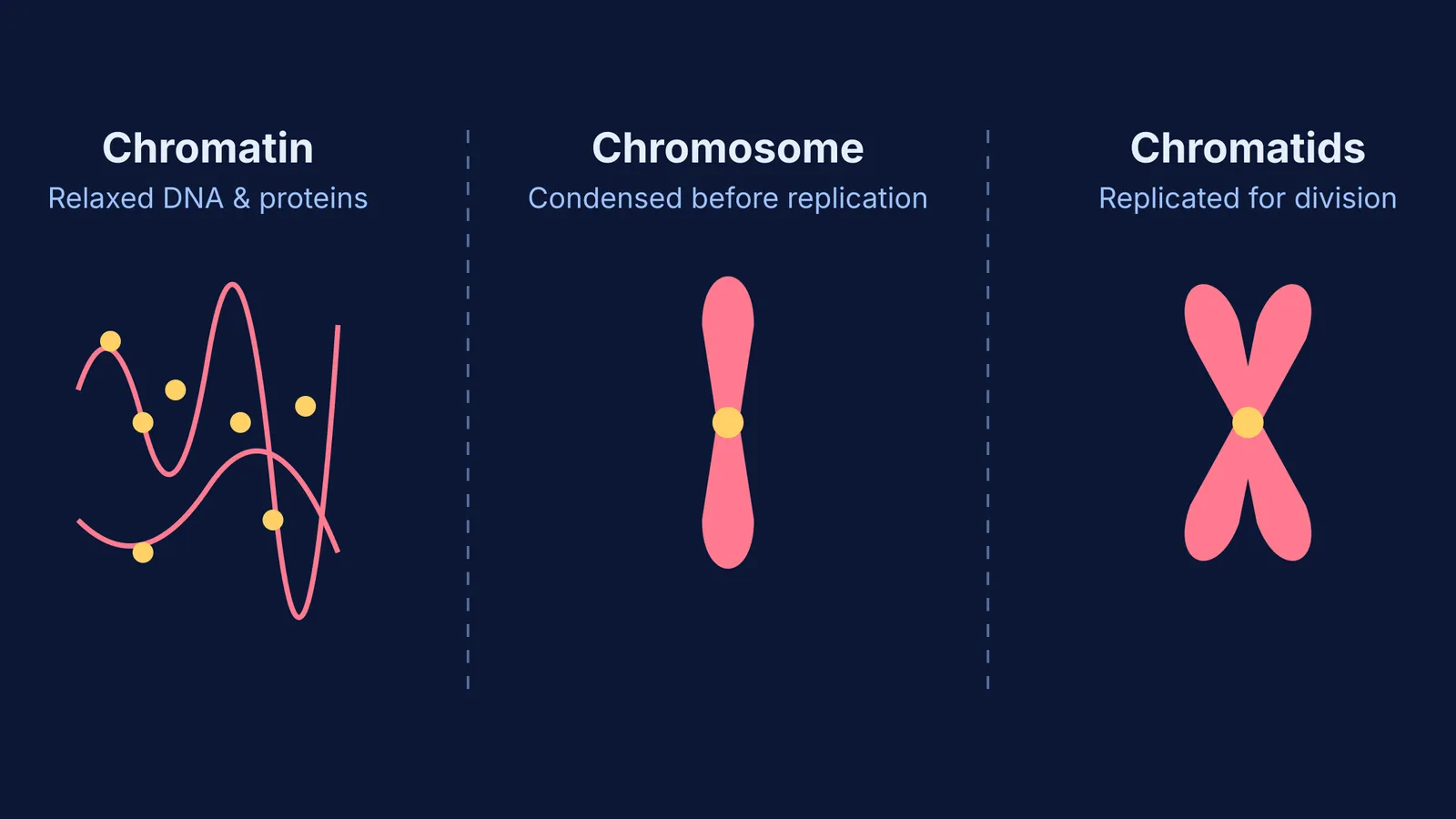
For most of a cell's life, it is not dividing. It is growing, metabolising and performing its standard functions. During this long resting phase (known as interphase), the genetic material inside the nucleus exists as chromatin.
According to the National Human Genome Research Institute, chromatin is a mixture of DNA and proteins that form the chromosomes found in the cells of humans and other higher organisms. The primary proteins involved are called histones. If you looked at chromatin under a powerful electron microscope, it would resemble a messy string of beads. The "string" is the DNA double helix, and the "beads" are the histone proteins that the DNA wraps around.
This relaxed, uncoiled structure is necessary. Because the DNA is spread out rather than tightly packed, the cell's machinery can easily access the genetic code. This accessibility allows the cell to read the genes to build necessary proteins (a process called transcription) and to copy the DNA completely before cell division (DNA replication).
What is a chromosome?
When a cell finally gets ready to divide, it faces a mechanical problem. It needs to move its DNA to opposite ends of the cell without tangling, tearing or breaking it. To solve this, the loose chromatin fibres coil up tightly into dense, robust structures.
Chromosomes are threadlike structures made of protein and a single molecule of DNA that serve to carry the genomic information from cell to cell. Without this tight packaging, the DNA contained in a single human cell would stretch to roughly 6 feet long. By supercoiling into chromosomes, the cell ensures the stable, safe storage and accurate segregation of its genetic material.
Humans typically have 23 distinct pairs of chromosomes, giving 46 in total. You inherit one half of each chromosomal pair from your mother and the matching half from your father.
What is a chromatid?
Before a cell can divide into two functioning new cells, it must make an exact copy of its DNA during the synthesis phase. Once this replication happens, the condensed chromosome takes on a familiar, symmetrical X-shape.
A chromatid is one of the two identical halves of a chromosome that has been replicated in preparation for cell division. These two halves are universally known as "sister chromatids," and they are joined together at a highly constricted central point called the centromere.
During the later stages of cell division, cellular machinery known as spindle fibres attaches to the centromere. The sister chromatids are then forcefully pulled apart to opposite ends of the dividing cell. Once they completely separate, each independent chromatid is considered its own individual chromosome in the newly formed daughter cells. If you map out the Cell cycle, mitosis and meiosis: stages and chromosome counts, you will see exactly when this critical split happens.
Seeing the difference with Fermi
The line between a chromosome and a chromatid often trips students up during biology exams, particularly when counting ploidy or DNA mass. It helps to map out the entire cell cycle visually rather than memorising text definitions in isolation. Fermi's Concept Library connects these terms directly to the stages of mitosis and meiosis, showing exactly when the DNA condenses, duplicates and separates.
If you are stuck on a genetics problem or cannot accurately picture how a cell divides, Fermi's AI tutor teaches rather than just gives you the answer. It can run a simulation of cell division, draw the dividing chromosomes on a board, and check your understanding as it goes. By working out what you already know, it builds a plan for the session so you sit down to a clear next step.


